
From calorimetry to medical imaging: a shining example of successful transfer!
A team at CERN has drawn inspiration from calorimetry methods developed for high-energy physics to create a new positron-emission tomography system for use in medical imaging, which they’ve dubbed AX-PET. With support from European and American laboratories*, the project is reaching fruition, as initial tests confirm its promise.

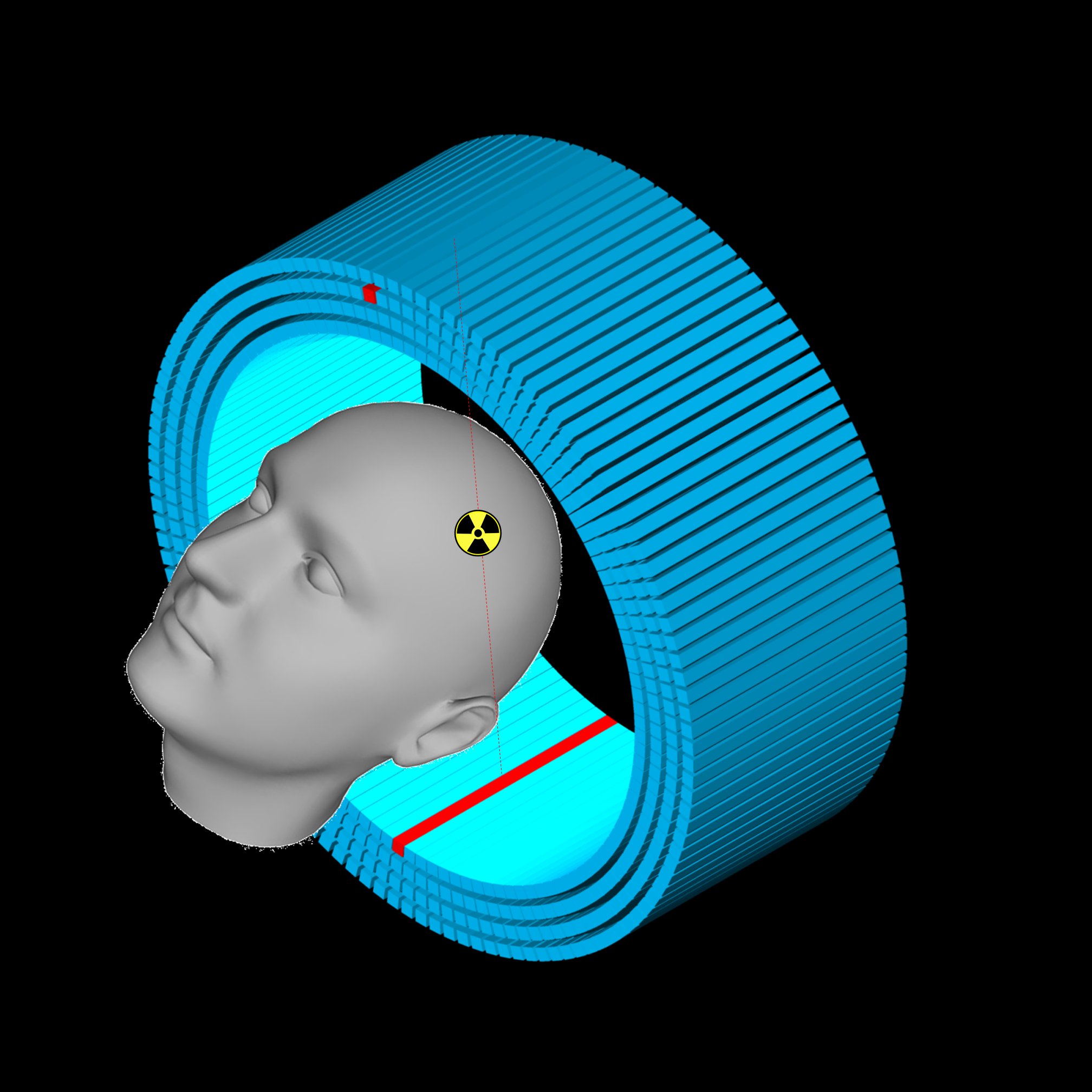
Snapshot of a “phantom”, a test object, surrounded by the AX-PET photon detectors.
Positron-emission tomography (PET) is a medical imaging technique based on the matter-antimatter interaction that can provide a three-dimensional representation of the metabolic activity of an organ. To do so, radioactive marker molecules are first injected into the subject. As the marker decays, it emits positrons (antimatter particles), which are annihilated upon encountering electrons in the surrounding environment. The resulting flash, consisting of two photons, is detected by the PET machine. In conventional PET systems, it is impossible to improve spatial resolution without losing sensitivity. AX-PET offers a way around this trade-off, improving the quality of the imaging. Thus, AX-PET can achieve millimetre-scale resolution, while conventional PET resolution is limited to 4 to 6 millimetres.
The AX-PET technique, whose development began in 2007, relies on calorimetry methods commonly used in particle physics: a lattice of crystals transforms the incoming photons into scintillating light. Each crystal is connected to a photodetector, which transmits analog information. The innovation in AX-PET compared to older technologies lies in the positioning of the crystals with respect to the photon source (see figure below). "Unlike the traditional layout, where the photosensitive crystals are positioned radially to the subject being examined, AX-PET uses elongated crystals, 100 mm in length, that are aligned parallel to the machine’s bore axis," explains Matthieu Heller, a Marie Curie fellow in CERN’s PH Department who is one of the names behind the project. "In this way, if you want to improve sensitivity, all you have to do is add more layers of crystals." Behind each row of crystals there is a perpendicular row of plastic strips, which is used to record the position at which a photon has impinged on the crystal. The resulting three-dimensional lattice can be used to measure the precise point of impact of a photon.
 |  |
| A traditional PET scanner showing the crystal layout. | The axial orientation of elongated crystals in AX-PET. |
In June the project team performed initial tests with small animals at the ETH Zurich laboratories that are specialised in small-animal imaging. "The results showed us that our demonstrator has reached the stage of maturity," notes Christian Joram, the leader of the PH Department’s Detector Technology Group and the person in charge of the AX-PET project. "There are now several possibilities. One of them would be to couple the PET machine with an MRI machine so as to give a combined snapshot of the metabolic and structural aspects of the subject. This would require some further work, because MRI—magnetic resonance imaging—relies on very strong magnetic fields, which must be protected against any interference from our detector."

Some members of the project team: Christian Joram (CERN), Chiara Casella (ETH Zürich) and Matthieu Heller (CERN) pose behind AX-PET.
Other avenues of research are aimed at improving the performance achieved by AX-PET, now that the demonstrator, which was manufactured at CERN, has shown that the new PET principle works and is feasible for bigger-scale application. These research ideas include simulations (using different crystals and geometries) and tests with new Digital SiPM photodetectors, which convert light directly into digital data. Coupled with the crystals, such detectors could be used to distinguish the time-of-flight of photons emitted by the source. This temporal information would improve the detection sensitivity and reduce the background noise, providing a much sharper image.
"As CERN researchers, our role isn’t to build a complete scanner, but to transfer the technology," concludes Christian Joram. "We have demonstrated the performance that is possible with our principle, and now we are helping our partners to develop suitable applications. Recently we have also started working with manufacturers and medical imaging experts on a detector that would incorporate both MRI and PET functionalities." From calorimetry to medical imaging, there is an unmistakable drive to move ahead!
*Laboratories involved in AX-PET project : INFN Bari, INFN Cagliari, INFN Roma, CERN, University of Michigan, Ohio State University, University of Oslo, Tampere University of Technology, IFIC Valencia, ETH Zürich.
by Caroline Duc